Home page

Diagnosis: Hodgkin's lymphoma

Česká verze

His good heart stopped beating on March 30, 2005.
Funeral will be held on April 2, 2005.

What is Hodgkin's lymphoma?

From the diary ...

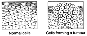
Hodgkin's lymphoma (HL) - Hodgkin's diseases is a malignant cell infiltration of the lymphatic system. The malignant cells clone or reproduce themselves so that in time the disease produces a partial or complete destruction of the normally healthy lymph tissue. As a result the lymph nodes become enlarged. The progression of the disease from its original site is due to lymph from the bodies' tissues circulating throughout the lymphatic system. The disease was first described by Thomas Hodgkin in 1832.

Hodgkin's Disease can present at any age. The age related incidence commonly occurs bimodally, with one peak young adults (20-30) and another after the age of fifty. Childhood Hodgkin's is rare. The male: female ratio is about 2:1. Nodular sclerosing is seen more often in young females. There may also be a genetic link to Hodgkin's, as there have been many reported incidences of family members of the same or different generation suffering from HD. To date the complete reasons as to why some people develop it and others don't remains largely unknown.

HL, like other cancers, are diseases of the body's cells. Cells in different parts of the body may work in different ways but all repair and reproduce themselves in the same way. Normally, this division of cells takes place in an orderly and controlled manner but if, for some reason, this process gets out of control the cells will continue to divide, developing into a lump or tumor.

Normal and tumour forming cells

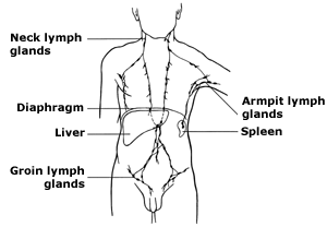
     The lymphatic system is one of the body's natural defences against infection. It is a complex system made up of  lymph organs, such as bone marrow, the thymus and the spleen, and lymph nodes which are connected by a network of tiny lymphatic vessels. Lymph nodes are found mainly in the neck, armpit and groin.

Lymphatic system

The malignant cells are thought to derive from dendritic reticulum cells, HD is identified almost exclusively by the presence of the large and binucleated Reed - Sternberg cells.

HD can be further sub classified in to four types. Classification can only be determined by histological examination, usually following a biopsy of the affected node. The type and the extent of the progression of disease, will then determine the prognosis and treatment plan; for instance those people that have Lymphocyte Predominance HD at stage 1a will have a higher remission rate than those people who have Lymphocyte Depletion at stage 4 b. Although, one must bear in mind that with so many medical advances it is fair to say that all types have a favourable prognosis. Statistically, over the past twenty years, the cure rate for HD has improved more than any cancer other than testicular cancer.

1- LYMPHOCYTE PREDOMINANCE

The histological appearance is largely of small cell lymphocyte proliferation, with only a few eosinophils, Reed - Sternberg cells and mononuclear Hodgkin's cells.

2- NODULAR SCLEROSING

This is the most common type. The histology shows the lymph node to be divided by broad bands of collagen or connective tissue, these bands encircle nodules containing a mixture of Reed - Sternburg cells, mononuclear Hodgkin cells, lymphocytes, plasma cells,, macrophages and eosinophills.

3- MIXED CELLULARITY

The lymph node is diffusely infiltrated with Reed - Sternburg cells, mononuclear Hodgkin cells, lymphocytes, plasma cells, macrophages and eosinphills. Fibrosis and focal necrosis are readily seen.

4- LYMPHOCYTE DEPLETION

There is either a reticular pattern, with a dominance of Reed - Sternburg cells, or a diffuse fibrosis where the lymph node is replaced by a disordered connective tissue, containing a few lymphocytes. Reed - Sternburg cells may also be infrequent.

The selection of the appropriate treatment depends on accurate staging of the extent of the disease. Your disease will be staged clinically, pathologically or both.
The clinical stage is determined by history, physical examination, x-rays, blood tests and your initial biopsy results.
The pathological stage is if involvement is found at laparotomy or by any further removal of tissue for histological examination other than that taken for the original diagnosis. Both clinical and pathological staging classification apply only to the time of disease presentation and prior to definitive therapy. However, pathological staging may be done during a course of treatment when symptoms demand that treatment to the upper half of the body must be given first, such as in mediastinal compression.
In most institutions the Ann Arbor staging model is used.

Stage I
Involvement of a single lymph node region (I) or of a single extra lymphatic organ or site (Ie).

Stage II
Involvement of two or more lymph node regions on the same side of the diaphragm (II) or localised involvement of extra lymphatic organ or site and of 1 or more lymph node regions on the same side of the diaphragm (Iie).

Stage III
Involvement of lymph node regions on both sides of the diaphragm (III) which may also be accompanied by localised involvement of extra lymphatic organ or site (IIIe) or by involvement of the spleen (IIIs) or both (IIIs e)

Stage IV
Diffuse or disseminated involvement of one or more extra lymphatic organs or tissues with or without associated lymph node enlargement. The reason for classifying at stage IV is identified further by defining sites by symbols.

N - Lymph nodes, S - Spleen, H - Liver, L - Lung, M - Marrow, O - Bone, P - Pleura, D - Skin

The systemic symptoms are further subdivided into A and B categories, B for those with defined general symptoms and A for those without.
The B classification is given to those people who have displayed:

(I) unexplained weight loss of more than 10% of the body weight in the six months before admission.
(II) unexplained fevers with temperatures above 38 st.C.
(III) night sweats.

Pruritis alone does not qualify for B classification.

Hodgkin's lymphoma treatment

  • Radiotherapy

  • Chemotherapy

  • Bone marrow transplantation (or peripheral blood stem cells transplantation)

Specific treatment depends on many factors including the type, and stage of lymphoma and the history and condition of the patient. Oncologists take these into account before recommending a treatment protocol.

Radiation therapy uses high-energy x-rays to kill cancer cells and shrink tumors. Radiation for Hodgkin's disease usually comes from a machine outside the body. It can be limited to a mantle field (such as the neck, chest or lymph nodes under the arm) or involve total nodal irradiation.

Chemotherapy uses drugs to kill cancer cells and shrink tumors. Chemotherapy can be taken by pill, or it may be taken intravenously. It is called a systemic treatment because the drugs enter the bloodstream and travel throughout the body. There are many different chemotherapy regimens used to fight lymphoma. Typical treatments for Hodgkin's include MOPP or ABVD or a combination of these drugs. Quite a few regimens exist for NHL, highly dependent on disease type.

Bone marrow (or stem cell) transplantation may be employed when the disease is resistant to other therapies. Because high doses of chemotherapy can destroy healthy bone marrow, the marrow can be replenished by 1) marrow removed from the body before treatment, frozen, then transfused back to the patient after chemotherapy (autologous transplants), or 2) marrow from a matched brother or sister or a marrow match from an unrelated donor (allogenic transplants).

Bone marrow is found inside the bones, particularly the pelvic bones. It is the `factory' for the blood, responsible for producing white blood cells (to protect against infection), red blood cells (to carry oxygen round the body) and platelets (to prevent bleeding).

Stem cells are blood cells at their very earliest stage of development in the bone marrow, before they have become committed to developing into white cells, red cells or platelets. It is these cells which are the key factor in transplants, whether just the stem cells are transplanted or the bone marrow itself (which naturally includes many stem cells).


... more about stem cells transplantation


Statistics

In the United States, about 7,400 new cases of Hodgkin's disease are expected this year (2000), with about 3,200 occurring in women and 4,200 occurring in men, according to the American Cancer Society (ACS).

The ACS lists lymphomas and leukemia together. In 1999, lymphomas and leukemia were the fifth most common type of cancer and the fourth leading cause of death by type of cancer.

About 1,400 people per year die from Hodgkin's disease in the United States, divided equally among males and females. However, death rates from Hodgkin's disease have fallen more than 60 percent since the early 1970s as more advances in treatment are made. More than 75 percent of people with Hodgkin's disease can be cured with radiation and/or chemotherapy.

The one-year survival rate after treatment is 93 percent. After five years, it is 82 percent, and after 10 years, it is 72 percent, according to the ACS. At 15 years, the overall survival rate is 63 percent. The most common cause of death in patients during the first 15 years after treatment is recurrent Hodgkin's disease. After 15 years, death is more likely due to other causes, such as a secondary type of cancer.


...

2.2.98 The Policka hospital

5.2.98 Computerized Tomography (CT scan) of the thorax - CT thorax (CT scan of the chest)

11.2.98 The Brno Faculty hospital.  The operation - cut a part of the tumour (about 50% tumour mass) from the mediastinum.

Histological exam.: H.lymphoma - nodular sclerosing type lymfocytedepletion (clinical stage II.B)

20.2.98 Starting VI cycles of ABVD chemotherapy (I.cycle)

21.2.98 CT scan of the abdomen - CT abdomen

9.3.98 2.chemo, I.cycle

23.3.98 3.chemo, II.cycle

6.4.98 4.chemo, II.cycle

20.4.98 5.chemo, III.cycle

22.-24.4.98 Implantation of venous port

4.5.98 6.chemo, III.cycle

18.5.98 7.chemo, IV.cycle

1.6.98 8.chemo, IV.cycle

12.6.98 CT thorax

15.6.98 9.chemo, V.cycle

1.7.98 10.chemo, V.cycle

14.7.98 11.chemo, VI.cycle

27.7.98 12.chemo, VI.cycle

8.9.98 CT thorax

9.9.98 CT abdomen

24.9.98 Radiotherapy planning - simulator

28.9.- 26.10.98 MOU Brno. Radiotherapy of the mediastinum - VARIAN2 (20 rations, 40 Gy)

14.12.98 CT thorax

13.2.99 CT abdomen

14.2.99 CT thorax

! RELAPS - the disease comes back !

25.2.-2.3.99 1.cycle of salvage chemotherapy VIM

15.3.99 The hair KO (first time)

25.-31.3.99 2.cycle -  VIM chemo

26.4.-2.5.99 3.cycle - VIM chemo

28.5.-10.6.99 Stimulation chemotherapy+separation of peripheral blood stem cells

19.7.99-17.8.99 High-grade chemotherapy+autologous transplantation peripheral blood stem cells

8.10.99 CT thorax

21.10.99 Radiotherapy planning- simulator

25.10.99- 22.11.99 Radiotherapy of the neck and above collar-bone - VARIAN1 (18 rations, 36 Gy)

24.2.00 CT thorax

30.3.00- 4.4.00 Remove of the venous port and broken katetr

20.2.01 CT thorax

!! RELAPS - the disease comes back again !!

6.3.01- 9.3.01 1.cycle of DHAP chemotherapy

14.3.01 CT abdomen

23.3.01- 29.3.01 Hospitalization for fever after chemotherapy DHAP

From the results of HLA typing of the sibling blood stem cells is not fitted for allogeneic transplantation peripheral blood stem cells.

9.4.01- 11.4.01 2. cycle - DHAP chemo

7.5.01- 9.5.01 3. cycle - DHAP chemo

30.5.01 CT thorax

18.6.01- 22.6.01 1.cycle of  Vepesid chemotherapy (oral)

16.7.01- 22.7.01 2.cycle - Vepesid chemo

13.8.01- 19.8.01 3.cycle - Vepesid chemo

5.9.01 CT thorax

20.9.01 TV report 1 (for playing-RealPlayer and min.28K modem required)

22.3.02 CT thorax

25.3.02 CT abdomen

!!! RELAPS - the disease comes back again !!!

2.4.02- 6.4.02 1.cycle of  CCNU+Vepesid+Dex.chemotherapy (P.O.)

1.5.02- 5.5.02 2.cycle CCNU+Vepesid+Dex.chemotherapy

28.5.02 TV report 2 (for playing-RealPlayer and min.28K modem required)

2.6.02- 6.6.02 3.cycle of CCNU+Vepesid+Dex.chemotherapy

26.6.02 CT thorax

16.8.02 CT thorax

10.9.02 Radiotherapy planning - simulator

16.9.02 - 10.10.02 Radiotherapy of the mediastinum - VARIAN2 (35 rations, 42 Gy)

10.1.03 CT thorax

!!! R E L A P S - the disease comes back - U.S.A. - paralysis of the legs!!!

13.6.03- 20.6.03 Inova Fairfax Hospital, Falls Church, VA, USA . CT, MRI, Dex.(I.V.).

13.9.03- 25.6.03 Radiotherapy of the neck spine area (C3-Th2) (10 days, 200 rad/day),

26.6.03- 8.7.03The Brno Faculty hospital, Czech Republic.  CT, MRI, Dex.(P.O.)

14.8.03- 9.9.03 The rehabilitation - The Brno Faculty hospital

23.9.03 CT thorax

24.9.03- 23.10.03 The rehabilitation - Jevíčko

10.10.03 MRI of the neck spine area

27.10.-29.10.03 1.cycle of BEACOPP chemotherapy

18.11.-24.11.03 Hospitalization for fever after chemotherapy - CT scan, X-Ray, US...

26.11.-28.11.03 2.cycle of BEACOPP chemotherapy

17.12.03 CT thorax

18.12.-20.12.03 3.cycle of BEACOPP chemotherapy

7.1.04 MRI of the neck spine area

8.1.-10.1.04 4.cycle of BEACOPP chemotherapy

10.2.04 PET scan (Positron Emission Tomography)

19.2.04 1.cycle of Gemzar chemotherapy (DEX, MSN, Durogesic...)

26.2.04 2.chemo, I.cycle

4.3.04 X-Ray of the neck spine area

10.3.04 3.chemo, I.cycle

1.4.04 1.chemo, II.cycle

8.4.04 2.chemo, II.cycle

10.4.-16.4.04 Hospitalization for fever after chemotherapy - X-Ray, Ultra Sound scan...

19.4.04 Blood infusion - 2 times

29.4.04 1.chemo, III.cycle

6.5.04 2.chemo, III.cycle

21.5.04 CT scan of the chest

26.5.04 CT scan of the neck

3.6.04 The implantation of the venous  port

8.6.-11.6.04 Hospitalization for the neck spine compression - DEX, X-Ray, Ultra Sound scan...

21.6.04 PET scan

30.6.-2.7.04 Hospitalization for the extraction of neck node sample

22.7.04 The day 1 - rituximab infusion

29.7.04 The day 8 - rituximab infusion + Zevalin radiolabelled [90Y]

15.9.-18.10.04 Hospitalization for fever - X-Ray, CT scan, B.lavage...

5.10.04 CT scan of the neck

7.10.04 CT scan of the chest

8.10.04 CT scan of the abdomen, remove of the venous port

16.10.04 PET scan

20.11.04-23.11.04 Vepesid 50 (2 days) chemotherapy +Dex.20mg (4 days) (oral)

3.12.04-6.12.04 Vepesid 100 (2 days) chemotherapy +Dex.20mg (4 days) (oral)

18.12.04-21.12.04 Vepesid 100 (2 days) chemotherapy +Dex.20mg (4 days) (oral)

1.1.05-4.1.05 Vepesid 100 (2 days) chemotherapy +Dex.20mg (4 days) (oral)

11.1.-4.2.05 Hospitalization for the progression of the disease - X-Ray, ATBs,  chemo...

12.1.05 HD Vepesid  chemotherapy (2mg/m2)

...

Allogenic bone marrow or peripheral blood stem cells transplantation

3.3.-6.3.05 Hospitalized at Brno University Hospital. Condition worsened, fatique, no appetite.


Main WWW links:

The Brno Faculty hospital

Cancer Bacup

Lymphoma Research Foundation of America

OncoLink


The page is still at work and it will, I hope so.

Thank you very much in advance for any kind of your help in this difficult situation.

Bank account: IBAN: CZ24 0800 0000 0006 1222 0143, BIC: GIBACZPX

Email: [email protected]


Last update: March 29, 2005

Encounters since 1/10/2000